Hospitals
CTG Interpretation in Labor: A Practical Guide
Aug 24 • 9 min read
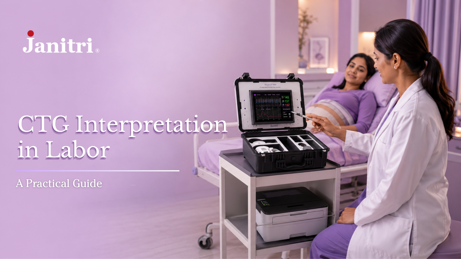
Table of Content
What Is CTG Monitoring?
How to Interpret a CTC
What Does DR C BRAVADO Stand For?
DR — Define Risk
C — Contractions
BRA — Baseline Fetal Heart Rate
110–160 bpm
Causes of fetal tachycardia can include:
Causes of fetal bradycardia can include:
V — Variability
A — Accelerations
D — Decelerations
Common Types of Decelerations
Early Decelerations
Variable Decelerations
Late Decelerations
Prolonged Decelerations
What Should You Do When a CTG Becomes Abnormal?
Stop or Reduce Oxytocin
Change Maternal Position
Check Maternal Oxygenation
Correct Maternal Hypotension or Dehydration
Examine the Mother
Consider Tocolysis
Umbilical Cord Blood Gases: Looking Beyond the CTG
CTG vs NST: What's the Difference?
The Future of CTG: Wireless Fetal Monitoring
Greater Maternal Mobility
Fewer Sensor Repositioning Issues
Centralized Monitoring
Remote Clinical Review
Key Takeaways for CTG Interpretation
Looking for CTG Monitoring Solutions for Your Hospital?
Conclusion
Frequently Asked Questions
1. What is CTG monitoring?
2. What is the normal fetal heart rate on CTG?
3. What is normal CTG variability?
4. What causes variable decelerations?
5. Do late decelerations mean fetal distress?
6. Does an abnormal CTG always mean a C-section?
7. Is CTG the same as NST?
About the CTG: Basics & Beyond Workshop
Cardiotocography (CTG) is one of the most widely used tools for monitoring fetal wellbeing during labor. It continuously records the fetal heart rate and uterine contractions, helping clinicians understand how the baby is responding to labor. Learn more about CTG machines, how they work, and their applications in hospitals.
But CTG interpretation isn't simply about looking at a line on a screen and deciding whether it is "normal" or "abnormal."
A fetal heart rate can change because of contractions, maternal blood pressure, medications, fetal sleep cycles, cord compression and several other factors. The challenge is understanding what the trace is telling you and what to do next.
These principles were discussed during the CTG: Basics & Beyond workshop, organized by Janitri in collaboration with the South Bangalore OBG-YN Society. The workshop brought together experienced clinicians, including Dr. Lata, Dr. Sri Lakshmi, Dr. Venuta, Dr. Veena, Dr. Bhargavi, Dr. Neelam and Dr. Srimati.
Here's a practical guide to the key principles of reading and interpreting a CTG trace during labor.
Clinical note: This article is for educational purposes and should not replace local protocols, clinical guidelines or professional judgment.
What Is CTG Monitoring?
CTG, or cardiotocography, continuously records the fetal heart rate and uterine contractions.
To interpret a CTG, clinicians assess several fetal heart rate features:
Baseline
Variability
Accelerations
Decelerations
Changes over time
These findings are interpreted alongside uterine activity, labor progress and the mother's clinical condition.
An important point to remember is:
An abnormal CTG does not automatically mean that the baby needs to be delivered immediately.
The first step is to understand what is happening, look for reversible causes and assess how the fetal heart rate responds to appropriate intervention.
How to Interpret a CTC
Trace: DR C BRAVADO
CTG traces can sometimes look complicated. A structured approach makes them easier to assess.
DR C BRAVADO is a commonly used framework for systematically reviewing a CTG trace during labor.
What Does DR C BRAVADO Stand For?
Term | What it means | What to assess |
DR – Define Risk | Understand the maternal and fetal risk factors before interpreting the trace. | Maternal condition, pregnancy risk, medications and reason for continuous monitoring. |
C – Contractions | Assess the frequency and duration of uterine contractions. | Number of contractions in 10 minutes, duration and signs of excessive uterine activity. |
BRA – Baseline Rate | Identify the average fetal heart rate between accelerations and decelerations. | Whether the baseline is within the expected 110–160 bpm range. |
V – Variability | Assess the small fluctuations around the fetal heart rate baseline. | Whether variability is absent, reduced, normal or increased. |
A – Accelerations | Look for temporary rises in fetal heart rate. | Their presence and relationship with fetal activity. |
D – Decelerations | Assess temporary falls in fetal heart rate. | Type, timing, duration, depth, recovery and relationship to contractions. |
O – Overall Assessment & Plan | Bring the CTG findings and clinical information together. | Overall pattern, possible causes, interventions, response and need for escalation. |
In simple terms: DR C BRAVADO helps clinicians move from clinical context → contractions → fetal heart rate → interpretation → action.
Let's look at each step in more detail.
DR — Define Risk
Before studying the trace, understand the clinical situation.
Ask:
Is this a low-risk or high-risk pregnancy? Why is continuous monitoring being used?
Important factors may include:
Maternal hypertension
Gestational diabetes
Maternal fever or infection
Previous uterine scar
Epidural analgesia
Fetal growth restriction
Meconium-stained liquor
The same CTG feature can have different significance depending on the clinical context.
C — Contractions
Look at uterine activity over a 10-minute period.
In active labor, around 3–5 contractions in 10 minutes is generally considered typical.
More than 5 contractions in 10 minutes is described as tachysystole.
Why does this matter?
When contractions occur too frequently, there may be less time for placental blood flow between contractions. If excessive contractions are accompanied by fetal heart rate abnormalities, the cause needs to be addressed promptly.
BRA — Baseline Fetal Heart Rate
The normal baseline fetal heart rate is generally:
110–160 bpm
A baseline above 160 bpm is considered fetal tachycardia, while a baseline below 110 bpm is fetal bradycardia.
Causes of fetal tachycardia can include:
Maternal fever or infection
Dehydration
Certain medications
Fetal anemia
Other causes of fetal compromise
Causes of fetal bradycardia can include:
Cord prolapse or compression
Placental abruption
Uterine rupture
Maternal hypotension
Dehydration
One simple but important check is to verify the maternal pulse. Occasionally, the monitor may be detecting the mother's heart rate instead of the fetus.
V — Variability
Variability refers to the small fluctuations in fetal heart rate around the baseline.
It is an important indicator of fetal autonomic nervous system activity.
Variability | Approximate range |
Absent | 0–2 bpm |
Reduced | 3–5 bpm |
Normal | 5–25 bpm |
Increased | >25 bpm |
Reduced variability does not automatically mean fetal hypoxia.
It can occur during fetal sleep and may also be associated with prematurity or certain medications.
The important questions are:
How long has the variability been reduced?
Are there other abnormal features on the CTG?
Persistent or worsening reduced variability, particularly when combined with other abnormalities, is more concerning.
A — Accelerations
An acceleration is a temporary rise in fetal heart rate.
For pregnancies at or beyond 32 weeks, a commonly used definition is:
At least 15 bpm above baseline for at least 15 seconds.
Accelerations are generally reassuring.
However, the absence of accelerations does not automatically mean that a CTG is abnormal if the other features remain reassuring.
D — Decelerations
Decelerations are temporary drops in fetal heart rate.
The important thing isn't simply seeing a drop. Clinicians consider:
When did it start?
How deep is it?
How long does it last?
How quickly does it recover?
What is happening with the contractions?
Is variability preserved?
Is the pattern becoming more frequent or severe?
Common Types of Decelerations
Type | Common cause | What to look for |
Early | Head compression | Usually gradual and linked with contractions |
Variable | Cord compression | Shape, duration and recovery |
Late | Reduced uteroplacental oxygen transfer | Timing relative to contractions |
Prolonged | Acute interruption of oxygenation | Duration and recovery |
Early Decelerations
Early decelerations are commonly associated with fetal head compression during labor.
They usually mirror the contraction, with the lowest fetal heart rate occurring around the peak of the contraction.
When they occur without other concerning features, they are generally considered physiological.
Variable Decelerations
Variable decelerations are commonly associated with umbilical cord compression.
Some variable decelerations are brief and recover quickly.
They become more concerning when they are:
Recurrent
Prolonged
Slow to recover
Associated with reduced variability
Increasing in frequency or severity
Late Decelerations
Late decelerations are commonly associated with reduced uteroplacental oxygen transfer.
They usually begin after the contraction has started and recover after the contraction ends.
Recurrent late decelerations, particularly when accompanied by reduced variability, require careful assessment.
Prolonged Decelerations
A prolonged fall in fetal heart rate can occur because of events such as:
Uterine hyperstimulation
Maternal hypotension
Cord compression
Cord prolapse
Placental abruption
Uterine rupture
This requires prompt clinical assessment to identify and address the underlying cause.
What Should You Do When a CTG Becomes Abnormal?
An abnormal CTG doesn't automatically mean immediate cesarean delivery.
The first step is to identify whether there is a reversible cause.
A useful approach is:
Assess → Identify the cause → Intervene → Reassess → Escalate if necessary
Depending on the situation, measures may include:
Stop or Reduce Oxytocin
If excessive uterine activity is contributing to fetal heart rate abnormalities, oxytocin may need to be stopped or reduced according to clinical protocol.
Change Maternal Position
Moving the mother to a lateral position can help improve maternal circulation and uteroplacental perfusion.
Check Maternal Oxygenation
Routine oxygen is not generally recommended for fetal heart rate abnormalities when the mother herself is not hypoxic. If maternal hypoxia is present, it should be treated.
Correct Maternal Hypotension or Dehydration
IV fluids may be appropriate when clinically indicated, with caution in women at risk of fluid overload.
Examine the Mother
Depending on the situation, assess for:
Cord prolapse
Placental abruption
Rapid labor progress
Uterine rupture
Other causes of acute fetal compromise
Consider Tocolysis
If excessive uterine activity continues alongside fetal heart rate abnormalities, acute tocolysis may be considered according to local protocol.
Umbilical Cord Blood Gases: Looking Beyond the CTG
CTG tells us what is happening during labor.
Umbilical cord blood gas analysis can provide additional information about the baby's acid-base status at birth.
Paired samples from the umbilical artery and vein can be particularly useful when there has been concern about intrapartum hypoxia.
The umbilical artery reflects the fetal acid-base state more directly, while the umbilical vein reflects blood returning from the placenta.
Cord gas results should always be interpreted alongside:
CTG findings
Apgar scores
Neonatal examination
Resuscitation requirements
Delivery circumstances
A low Apgar score alone does not prove intrapartum hypoxic injury.
CTG vs NST: What's the Difference?
CTG and NST are related but serve different purposes.
CTG | NST |
Records fetal heart rate and uterine activity | Primarily assesses fetal heart rate reactivity |
Commonly used for intrapartum monitoring | Commonly used for antenatal surveillance |
Looks at baseline, variability, accelerations and decelerations alongside contractions | Focuses particularly on fetal heart rate response |
In simple terms:
NST mainly evaluates fetal heart rate reactivity, while CTG combines fetal heart rate monitoring with uterine activity.
For a detailed comparison, see our guide to CTG vs NST.
The Future of CTG: Wireless Fetal Monitoring
Traditional CTG has transformed intrapartum monitoring, but it also has practical limitations.
Conventional sensors can:
Lose signal
Require repositioning
Become difficult to maintain as the baby descends
Restrict maternal movement
Newer technologies are exploring transabdominal fetal ECG and EMG-based uterine activity monitoring.
Wireless systems can potentially provide:
Greater Maternal Mobility
Women may be able to move more freely during labor while remaining monitored.
Fewer Sensor Repositioning Issues
Alternative signal acquisition can help address some limitations associated with conventional transducers.
Centralized Monitoring
Data can be transmitted to a central monitoring system so that the clinical team can review multiple laboring patients.
Remote Clinical Review
Where supported by the technology and clinical workflow, senior clinicians may be able to review traces remotely.
The goal isn't simply to make monitoring wireless.
The goal is to make fetal monitoring more reliable, practical and clinically useful.
Key Takeaways for CTG Interpretation
When reading a CTG, don't focus on one feature.
Work through the trace systematically:
1. Define the risk
Understand the mother, fetus and clinical situation.
2. Check contractions
Look for excessive uterine activity.
3. Check the baseline
Normal is generally 110–160 bpm.
4. Assess variability
Look at the overall pattern and duration of any reduction.
5. Look for accelerations
Their presence is reassuring, but absence alone isn't necessarily abnormal.
6. Analyze decelerations
Ask what type they are and why they may be happening.
7. Look for reversible causes
Maternal hypotension, excessive contractions and cord problems may be treatable.
8. Reassess after intervention
Don't just act—look at how the fetal heart rate responds.
Looking for CTG Monitoring Solutions for Your Hospital?
Explore Janitri's fetal monitoring solutions designed to support CTG monitoring and modern maternity care workflows.
[Explore CTG Monitoring Solutions]
Conclusion
A CTG trace is more than a set of lines on a monitor.
Good CTG interpretation means combining the fetal heart rate, contractions and clinical situation to understand what is happening to the baby.
The DR C BRAVADO framework provides a practical way to approach each trace:
Define Risk → Contractions → Baseline → Variability → Accelerations → Decelerations → Overall Assessment
And when the trace becomes concerning, the question shouldn't immediately be:
"Do we need to deliver?"
Instead, start with:
"What is happening, what could be causing it, can we correct it, and how is the baby responding?"
That approach can help clinicians make more informed decisions while avoiding unnecessary intervention whenever the clinical situation allows.
Frequently Asked Questions
1. What is CTG monitoring?
CTG, or cardiotocography, continuously monitors fetal heart rate and uterine contractions, particularly during labor when continuous electronic fetal monitoring is clinically indicated.Learn more about CTG machines and fetal monitoring used in hospital settings.
2. What is the normal fetal heart rate on CTG?
The commonly accepted normal baseline fetal heart rate is 110–160 bpm.
3. What is normal CTG variability?
Normal baseline variability is generally 5–25 bpm.
4. What causes variable decelerations?
Variable decelerations are commonly caused by umbilical cord compression. Their clinical significance depends on their duration, frequency, recovery and associated CTG features.
5. Do late decelerations mean fetal distress?
Late decelerations can indicate reduced uteroplacental oxygen transfer and require assessment, particularly when they are recurrent or associated with other abnormal CTG features.
6. Does an abnormal CTG always mean a C-section?
No. The team should first assess the clinical situation, identify reversible causes, intervene where appropriate and reassess. The mode and timing of delivery depend on the complete clinical picture.
7. Is CTG the same as NST?
No. NST primarily evaluates fetal heart rate reactivity, while CTG records fetal heart rate together with uterine activity.
About the CTG: Basics & Beyond Workshop
This article is based on the clinical discussions from the CTG: Basics & Beyond workshop, organized by Janitri in collaboration with the South Bangalore OBG-YN Society.
The workshop brought clinicians together to discuss practical CTG interpretation, fetal monitoring, decelerations, intrauterine resuscitation, cord blood gas analysis and emerging fetal monitoring technologies.
Clinical contributors included: Dr. Lata, Dr. Sri Lakshmi, Dr. Venuta, Dr. Veena, Dr. Bhargavi, Dr. Neelam and Dr. Srimati.
Medical disclaimer: This article is for educational purposes and does not replace professional medical advice, local hospital protocols or current clinical guidelines.