Hospitals
Breech Baby Position: Signs, Risks & What You Can Do
Apr 30 • 5 min read
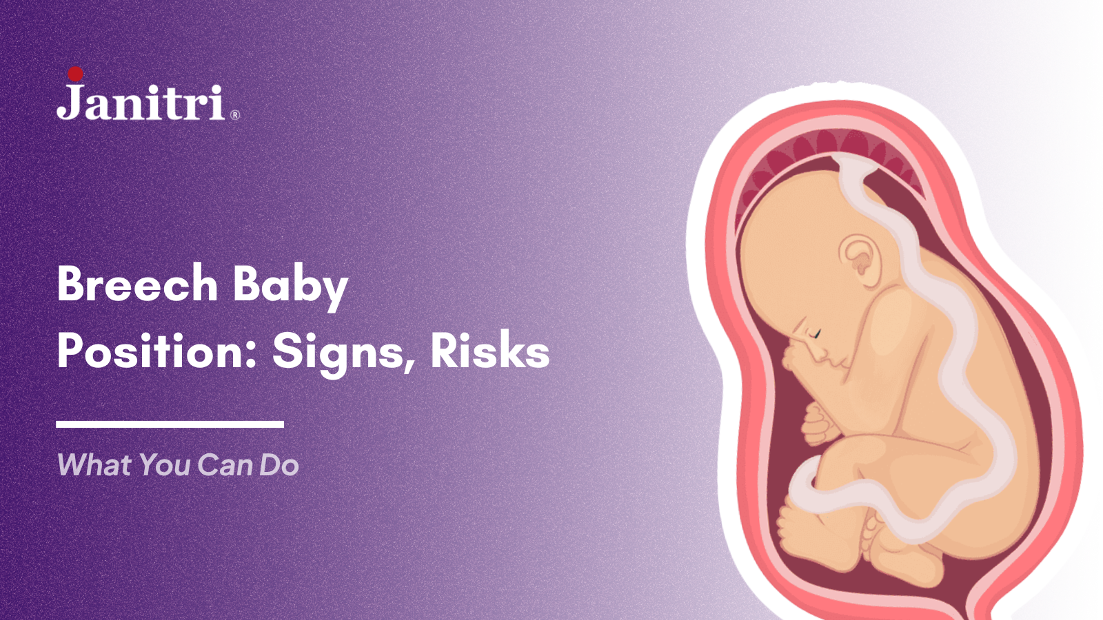
Table of Content
What Is the Breech Baby Position?
Types of Breech Baby Position
Signs of Breech Baby Position
Why Breech Presentation Occurs?
1. When is breech baby position considered a concern?
2. Are clinical signs enough to diagnose breech position?
3. Is vaginal delivery safe in breech cases?
4. What is the role of ECV in breech management?
5. Why is continuous monitoring important in breech cases?
In a busy labor room or during a routine third trimester scan, identifying a breech baby position is rarely just a positional finding. It becomes a clinical decision point that influences monitoring strategy, delivery planning, and team preparedness.
For obstetricians and care teams, breech presentation demands a balance between evidence-based protocols and individualized judgment. The question is not only about fetal position, but about timing, safety, and the ability to respond to evolving intrapartum scenarios.
This article explores the signs of breech baby position, associated risks, and practical management pathways, with a focus on real-world clinical application.
What Is the Breech Baby Position?
A breech baby position in the womb refers to a fetus presenting with the buttocks or feet closest to the birth canal instead of the head. While common earlier in pregnancy, most fetuses achieve cephalic presentation by 36 to 37 weeks.
Persistent breech presentation at term is clinically significant as it directly impacts delivery planning. It requires careful assessment of fetal condition, maternal factors, and institutional readiness.
Types of Breech Baby Position
Understanding the type of breech is essential for risk stratification and delivery decisions.
Type of Breech | Description | Clinical Relevance |
Frank Breech | Hips flexed, knees extended, buttocks presenting | Most common and relatively more favorable for vaginal delivery in selected cases |
Complete Breech | Hips and knees flexed, fetus in a sitting posture | Less common and associated with variable outcomes |
Footling Breech | One or both feet presenting first | Higher risk of cord prolapse and usually not preferred for vaginal delivery |
Before interpreting the table, it is important to note that not all breech presentations carry equal risk. The type of breech, combined with fetal size and maternal pelvic assessment, plays a critical role in determining the safest delivery approach. Clinical decisions should therefore be individualized rather than protocol-driven alone.
Signs of Breech Baby Position
Clinical suspicion of signs of breech baby position often arises during abdominal examination and patient-reported fetal movements.
Fetal head palpable in the fundus and a softer mass in the lower abdomen may suggest breech presentation. This requires confirmation through ultrasound before any clinical decisions are made.
Patients may report stronger kicks in the lower abdomen and less pressure in the pelvis. These observations are subjective and should not be used for diagnosis.
Mothers often rely on fetal movement tracking at home to notice unusual patterns before clinical confirmation.
On auscultation, fetal heart sounds may be heard above the umbilicus. This can guide suspicion but is not definitive without imaging support.
Why Breech Presentation Occurs?
In many cases, breech presentation has no identifiable cause and should not be attributed to modifiable maternal factors.
Prematurity is one of the most common reasons, as fetal mobility is higher and spontaneous version may still occur.
Placental location, especially fundal or previa, can influence fetal positioning by altering available space.
These factors are more commonly assessed during pregnancy trimesters changes, especially in late second and third trimester.
Uterine anomalies or altered amniotic fluid volume can restrict or facilitate fetal movement, impacting final presentation.
Risks of Breech Baby
The risks of breech baby are primarily associated with delivery rather than antenatal development, but they require careful anticipation.
Cord prolapse is a significant concern, especially in footling breech, where presenting parts do not adequately fill the pelvic inlet. This can lead to acute fetal compromise.
Early detection through fetal distress monitoring is critical in high-risk labour scenarios.
Head entrapment during vaginal delivery is a critical risk, particularly in preterm fetuses where the head is proportionally larger. This necessitates experienced handling.
Intrapartum fetal distress may occur due to compression or delayed delivery, reinforcing the importance of continuous fetal monitoring.
What You Can Do If a Baby Is Breech?
Management of a breech baby position should focus on evidence-based interventions and structured monitoring.
Expectant management may be appropriate before 36 weeks, as spontaneous version remains possible. Regular follow-up ensures timely reassessment.
External Cephalic Version can be considered in eligible patients under controlled settings. Success depends on multiple factors including parity, fluid levels, and placental position.
Continuous fetal monitoring plays a crucial role in detecting early signs of compromise. Integrating real-time monitoring systems can improve clinical response and decision-making.
Delivery Options for Breech Baby
Delivery planning requires a case-by-case evaluation rather than a uniform approach.
Vaginal breech delivery may be considered in carefully selected patients where clinical criteria are met and experienced personnel are available. Institutional protocols and readiness are key.
Planned cesarean section remains the preferred option in many settings, particularly for footling breech or when risk factors are present. It offers a more controlled delivery environment.
Clinical Considerations at a Glance
Aspect | Key Consideration |
Diagnosis | Confirm with ultrasound, not clinical suspicion alone |
Monitoring | Continuous fetal monitoring improves safety during labor |
Intervention | ECV may be offered in selected cases |
Delivery Planning | Individualized based on type of breech and maternal factors |
Risk Management | Preparedness for emergency intervention is essential |
Before applying these considerations, it is important to recognize that breech management is highly context-dependent. The availability of skilled personnel, monitoring infrastructure, and institutional protocols significantly influences outcomes. Standardization helps, but flexibility in clinical judgment remains equally important.
The Role of Monitoring in Breech Management
Breech presentation places increased emphasis on fetal surveillance, particularly during labor. Subtle variations in fetal heart rate patterns can indicate early compromise.
Continuous monitoring systems enable clinicians to track these changes more effectively, reducing reliance on intermittent assessments. In high-risk scenarios such as breech presentation, this can contribute to more timely interventions and improved outcomes.
Conclusion
A breech baby position is not an uncommon finding, but it requires a structured and thoughtful approach to management. The focus should remain on accurate diagnosis, risk stratification, and ensuring the safest possible delivery pathway.
For clinicians, the challenge lies not just in identifying breech presentation, but in navigating the decisions that follow with clarity and preparedness. With appropriate monitoring, skilled care, and patient-specific planning, breech cases can be managed with confidence and safety.
FAQs
1. When is breech baby position considered a concern?
Breech presentation becomes clinically significant after 36 to 37 weeks when spontaneous version is less likely. At this stage, delivery planning and monitoring strategies need to be defined.
2. Are clinical signs enough to diagnose breech position?
No, clinical signs can raise suspicion but are not definitive. Ultrasound remains the gold standard for confirming fetal presentation.
3. Is vaginal delivery safe in breech cases?
It can be safe in selected cases with strict criteria and experienced clinicians. However, many cases are managed with planned cesarean section for better control.
4. What is the role of ECV in breech management?
External Cephalic Version is a recommended option for eligible patients. It can reduce the need for cesarean delivery if successful.
5. Why is continuous monitoring important in breech cases?
Breech presentation carries a higher risk of intrapartum complications. Continuous monitoring helps detect early signs of fetal distress and enables timely intervention.